Eyecare
Eye Care Services
With a full range of eye health services, our team of experts are dedicated to helping you enjoy healthy eyes and sharp vision. Acton’s Eye Care Centre offers a full range of services to keep you healthy and happy! Whether it’s a new pair of eyeglasses or treatment for an eye disease, one call can get everything taken care of! We cater to all ages so come see us for all your eye care needs!
Eye Exams, Eye Emergencies & More
Acton Eyecare is more than just an eye doctor’s office. We provide prescriptions for glasses and contact lenses, corrective laser surgery (including LASIK), as well as comprehensive exams to help you reach your full potential vision-wise! We’re always here for you! Call us today to set up an appointment and find out more about how we can help.
Comprehensive Eye Exams
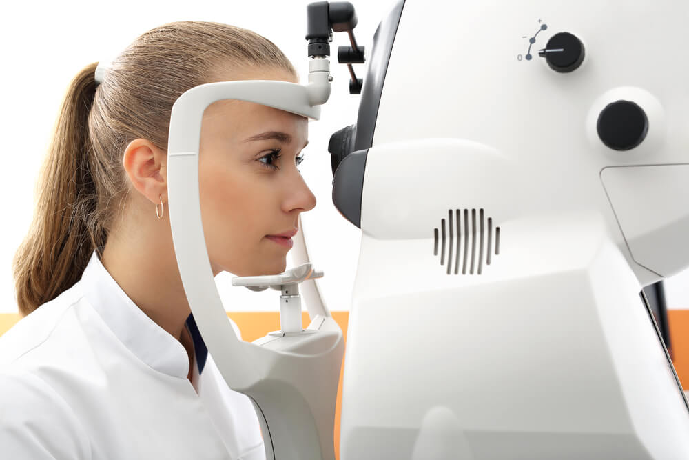
It’s never too early or late to start thinking about your eyes. Your eyesight is not only important for what we see, but also as an indicator of our overall health.. A comprehensive eye exam includes a number of tests and procedures that will examine the health, quality & functionality (or lack thereof) for both eyes.
Your eye doctor will not only determine the prescription for eyeglasses or contact lenses, but also check the health of your eyes and vision. A comprehensive exam includes tests that range from simple ones like having you read an eye chart to more complex procedures such as using a high powered lens inside the socket known as ‘the cup’. A good rule? Our doctors recommend completing an eye examination every one-three years depending on age group!
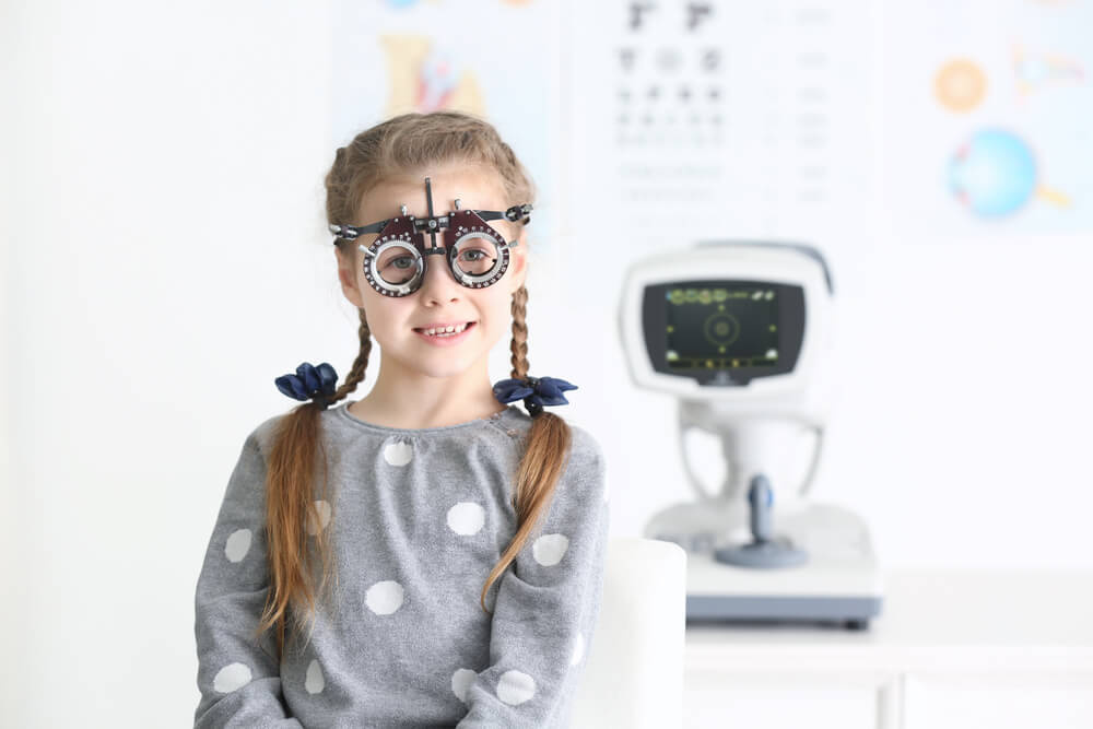
Eye Exams for Children
The Canadian Association of Optometrists (CAO) recommends that all children between the ages of 6-9 months should have their eyes examined again before they start school, as well at 2 and 5 years old. School aged children should see an optometrist every year throughout their schooling career, from grade 1 until college or university graduation.
- Premature birth
- Developmental delays
- Crossed or turned eyes
- Genetics predisposition to eye disease
- Past eye injury
- Other diseases
The CAO recommends that you should have your child’s eyes examined at least once every 12 months or according to your optometrist’s instructions.
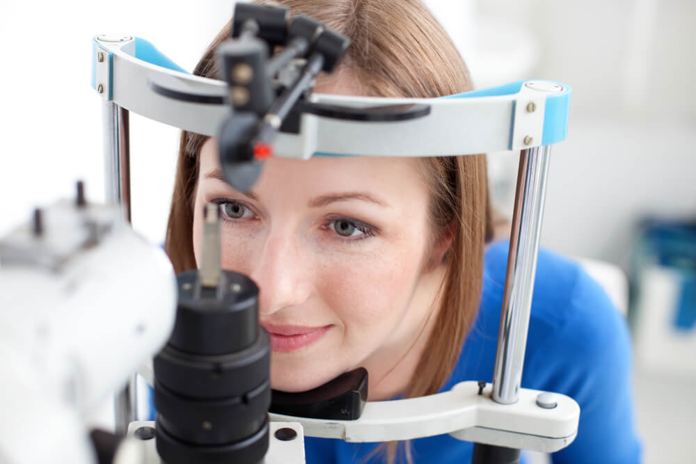
Eye Exams for Adults
The CAO also recommends an annual eye exam for any adult who wears eyeglasses or contacts. If you don’t normally need vision correction, you still need an eye exam every two to three years up to the age of 40, depending on your rate of visual change and overall health. Doctors often recommend more frequent examinations for adults with diabetes, high blood pressure and other disorders, because many diseases can have an impact on vision and eye health.
If you are over 40, it’s a good idea to have your eyes examined every one to two years to check for common age-related eye problems such as presbyopia, cataracts and macular degeneration.
Because the risk of eye disease continues to increase with advancing age, everyone over the age of 50 should be examined annually.
Advanced Eye Care
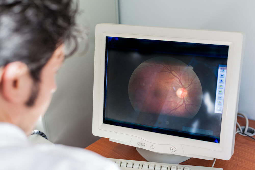
In order to provide the best possible care for our patients, we use cutting-edge digital imaging technology that will not only allow for visual assessments but an electronic record of your retina. With this technology, we are able to assess your eyes and detect diseases in its early stages. If conditions are detected at an appropriate point of time. it may be treated successfully without any loss of vision.
The eye doctor will store your retina’s images electronically so that he or she can check for changes in the future. This is important because many conditions, such as glaucoma and macular degeneration are diagnosed by noticing changes over time. The use of retinal images stored electronically provides doctors more knowledge about how well treatments are working based on consistent monitoring or if any alternatives need to be explored. As mentioned, many types of diseases such as glaucoma, and macular degeneration have been found through regular tests performed every few years.
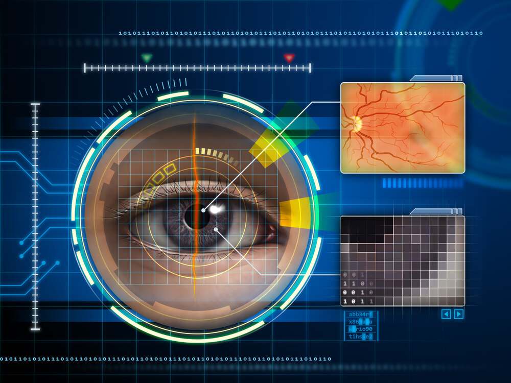
Digital Retinal Imaging
Digital retinal imaging is a new technology that takes an accurate digital photo of your retina. This imaging provides an accurate assessment into the overall health of your eyes. Digital retinal imaging is able to provide valuable information about any future examinations and detect whether there has been any changes in your vision due illnesses or trauma over time.
The advantages of digital imaging include:
- Utilizes eye-safe near-infra-red light
- Image resolution is extremely high quality
- Rapid, safe, non-invasive and painless
- No patient prep required
- Delivers detailed images of your retina and sub-surface of your eyes
- Delivers instant, direct imaging of the form and structure of eye tissue
Optical Coherence Tomography (OCT)
An Optical Coherence Tomography scan (commonly referred to as an OCT scan) is the latest advancement in imaging technology. Similar to ultrasound, this diagnostic technique employs light rather than sound waves to achieve higher resolution pictures of the structural layers of the back of the eye.
A scanning laser used to analyze the layers of the retina and optic nerve for any signs of eye disease, similar to an CT scan of the eye. It works using light without radiation, and is essential for early diagnosis of glaucoma, macular degeneration and diabetic retinal disease.
With an OCT scan, doctors are provided with colour-coded, cross-sectional images of the retina. These detailed images are revolutionizing early detection and treatment of eye conditions such as wet and dry age-related macular degeneration, glaucoma, retinal detachment and diabetic retinopathy.
An OCT scan is a noninvasive, painless test. It is performed in about 10 minutes right in our office. Feel free to contact our office to inquire about an OCT at your next appointment.
Visual Field Testing
A visual field test measures the range of your peripheral or “side” vision to assess whether you have any blind spots (scotomas), peripheral vision loss or visual field abnormalities. It is a straightforward and painless test that does not involve eye drops but does involve the patient’s ability to understand and follow instructions.
- Digital Retinal Imaging
Used to detect diseases such as diabetes and macular degeneration, it is critical to confirming the health of the retina, optic nerve and other retinal structures. - Visual Field Testing
Measures the quality of your side vision (peripheral vision). Usually involves covering one eye and focusing the other on a fixed point in front of you, while describing what you can see on the “periphery” of your vision. - Glaucoma Testing
Glaucoma testing involves measuring internal eye pressure and a detailed scan of the retina for signs of disease.
LASIK Surgery
LASIK is a popular vision-correcting procedure that can be done at our clinic. Our doctors have extensive experience with the preoperative evaluation and post surgical care of LASIK eye surgery.
There are other options as well that are just as effective. We are able to find an excellent solution for you by working closely together to find out what procedure best suits your needs. Get in contact with our team today and start your consultation!
Introduction to LASIK
LASIK is the most popular refractive surgery procedure. The name of this surgery is called “laser-assisted in situ keratomileusis” which in short-form is pronounced LASIK. Another commonly used term to refer to this procedure is called “LASIX”.
LASIK surgery has become quite a popular vision procedure because it has advantages over other procedures. Some of these advantages include few or no side effects, and quick recovery time. Patients usually achieve good eyesight within one day.
In LASIK eye surgery, a microkeratome is used to create a thin and circular flap in your cornea. This instrument has been around for quite some time but newer techniques have made it possible that we can now do this with laser cuts instead.
The excimer laser is used to remove tiny bits of tissue from the cornea, this in turn allows it to be reshaped perfectly. When the surgeon removes some corneal tissue underneath using an excimer laser, it is done so with pinpoint accuracy. The cool ultraviolet light beam created by this device assists with precisely removing the (“ablate”) tissue.
When the cornea is reshaped, it is done so in a way that makes it better for focusing light into your eye and onto the retina. This will allow the patient to have clearer vision than before. After this procedure is completed, the flap is then laid back over where the removal took place.
The LASIK procedure is a great option for both nearsighted and farsighted people. With nearsighted people, the goal is to flatten out a too-steep cornea. The goal with farsighted individuals is to create steeper angles. Excimer lasers are capable of correcting astigmatism by smoothing an irregular surface into something more normal in shape.